In the ever-evolving world of medicine, advancements in technology have significantly transformed the way we diagnose and understand diseases. One such innovation that has reshaped the field is digital imaging in macro pathology.
The days of traditional gross examination are over; today, pathologists can capture high-resolution images of entire specimens and study them in minute detail on computer screens. But before we delve into the details, let’s establish the basics.
What is Macro Pathology?
Macro pathology, often referred to as gross pathology, is a branch of pathology that involves the examination of diseases with the naked eye. In this context, ‘gross’ doesn’t necessarily mean unpleasant; it’s derived from the Latin ‘grossus,’ meaning large. So, macro pathology is all about studying the larger, visible aspects of the disease.
Pathologists in this field examine organs, tissues, and bodily fluids to diagnose diseases. They look at the color, size, shape, and texture of specimens to identify abnormalities. It’s a bit like being a detective, but instead of solving crimes, pathologists are unraveling the mysteries of diseases. They’re the Sherlock Holmes of the medical world, using their keen observation skills to make diagnoses and guide treatment.
What is Digital Imaging?
Digital imaging is a process that uses digital technology to capture, store, manipulate, and display images. In simple terms, it’s like taking photos with a high-tech camera. But instead of just snapping selfies or picturesque landscapes, digital imaging in medicine captures images of tissues, cells, and other biological structures.
Digital imaging has revolutionized many fields, from photography to astronomy, and medicine is no exception. It allows us to see things in ways we never could before, offering a level of detail and precision that was once unimaginable.
How Can Digital Imaging Be Used in Macro Pathology?
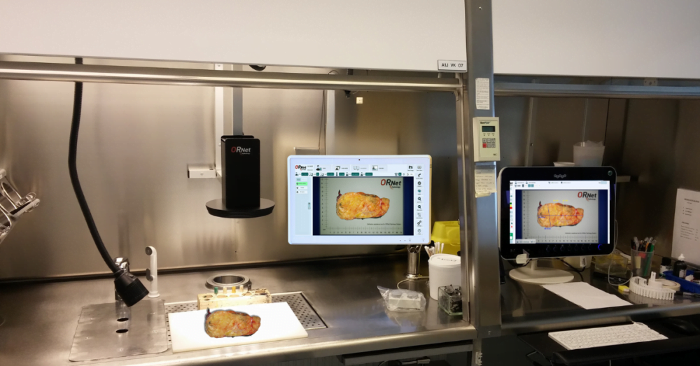
Now, here’s where things get really interesting. By combining digital imaging with macro pathology, we can take detailed pictures of specimens, magnify them, and analyze them with incredible precision. It’s like having a super-powered microscope but for the grossing process instead of examining glass slides.
Imagine being able to zoom in on a tissue sample to see the tiniest details, or to enhance an image to make certain features stand out. With digital imaging, pathologists can do all this and more. It’s a game-changer for disease diagnosis and research.
Benefits of Digital Imaging in Macro Pathology
The fusion of digital imaging with macro pathology is comparable to a powerful alliance, each contributing its distinct strengths to forge something truly remarkable. Let’s delve into why this combination is causing a stir in the medical field:
1. Improved Documentation:
Digital images provide a permanent, unchanging record of the macroscopic appearance of a specimen. This is invaluable for documentation purposes. It’s like having a photographic memory that never fades, ensuring that every detail is captured and preserved.
Instead of relying on verbal cues, pathologists can actually see what the specimen looked like before dissection. Also, every image captured serves as irrefutable proof. This adds traceability to the gross processing step and increases patient security.
2. Increased Accuracy:
Digital imaging isn’t just about taking high-quality pictures for storage; it’s a tool for analysis. These images can be magnified, enhanced, and manipulated, helping pathologists to identify subtle features that may be missed by the naked eye. It’s like having a magnifying glass that can zoom in on the tiniest details, revealing things that were previously hidden. This can lead to more accurate diagnoses, better patient care, and new insights into diseases.
3. Reduced Turnaround Time:
In medicine, time is often of the essence. The quicker a diagnosis can be made, the sooner treatment can begin. Digital imaging can help to speed up this process. Images can be captured, stored, and analyzed quickly, reducing the time it takes to produce a diagnostic report.
4. Improved Communication:
One of the great things about digital images is that they can be easily shared. This can improve communication and collaboration between pathologists and other healthcare professionals. In other words, It’s like having a group chat for medical professionals, where everyone can see the same images and discuss their findings.
Instead of manual sharing, some gross pathology imaging solution providers include consultation and sharing features that make the workflow more efficient.
5. Enhanced Training:
Digital images are an excellent resource for education. They can be used to train new pathologists, providing them with a wealth of real-world examples to study. They can also be used for continuing education, helping experienced pathologists to keep their skills sharp and stay up-to-date with the latest developments in the field.
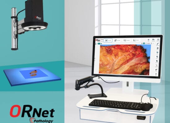
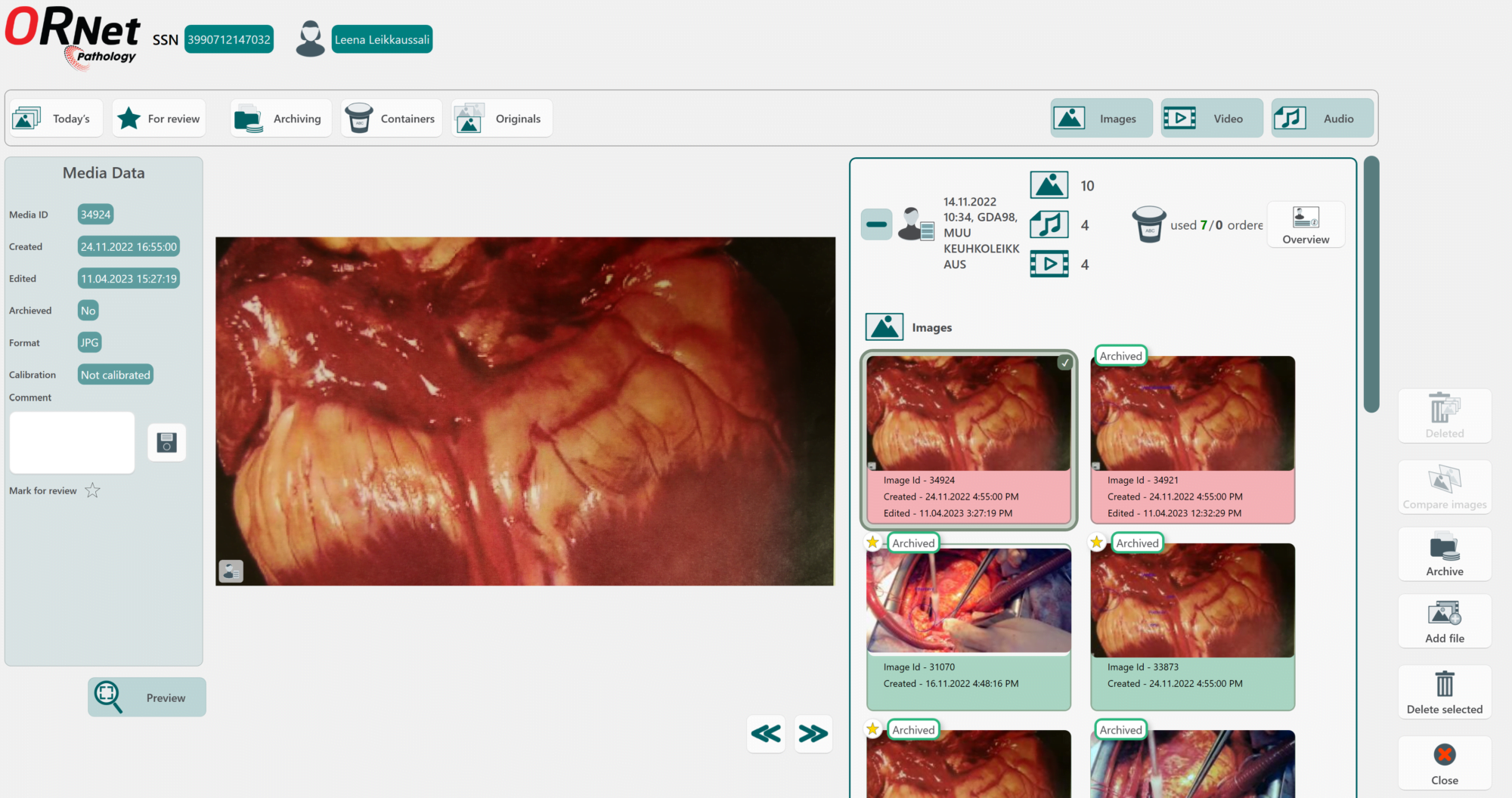
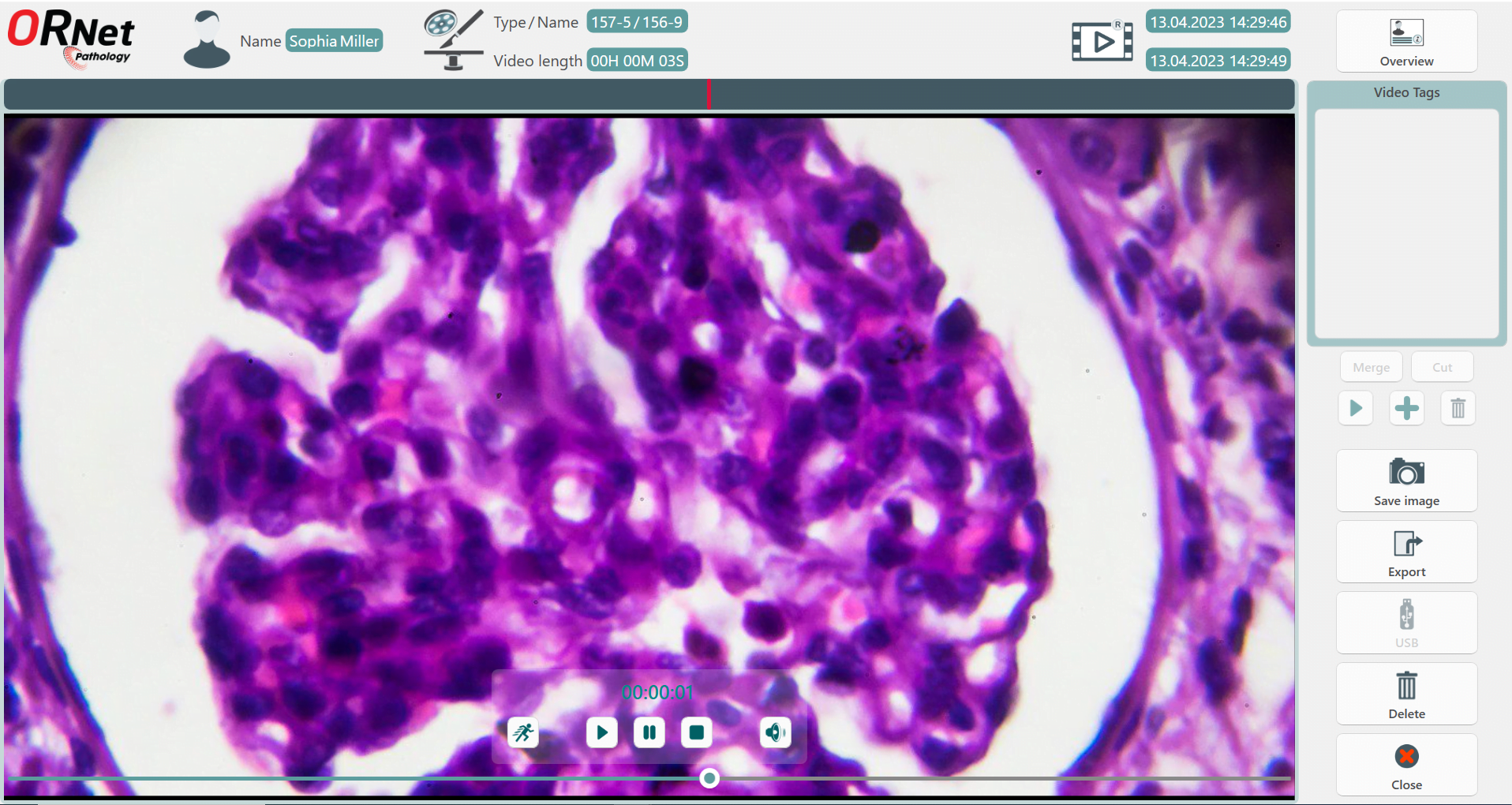
For that purpose, ORNet Pathology offers 4K video recording with a built-in video editor function. This can be used to cut important parts from the video and prepare precise training materials.
6. Increased Research Opportunities:
Research is the backbone of medical advancement, and digital images open new doors for exploring diseases. With a wealth of data at their fingertips, researchers can study the macroscopic appearance of diseases in unprecedented detail. As a result, researchers can obtain new insights, improved diagnostic methods, and even new treatments.
Challenges of Digital Imaging in Macro Pathology
Of course, every technological advancement comes with its own challenges, and the integration of digital imaging into macro pathology is no exception. Here are some of the hurdles that need to be overcome:
I. Cost:
High-tech gadgets, including digital imaging systems, often carry a hefty price tag, reflecting the advanced technology they encompass. The cost of these systems can be a significant barrier for some laboratories, particularly those with limited budgets. However, as technology advances and becomes more widespread, the cost of digital imaging systems is likely to decrease.
II. Technical Expertise:
Digital imaging systems are sophisticated pieces of technology. And using them requires a certain level of technical expertise. This means that laboratories need to hire new people and/or invest in training for their staff to ensure they can maintain and use these systems effectively. As a result, their costs go up.
III. Data Security:
Digital images contain sensitive patient data, so it’s crucial to have robust security measures in place. This includes secure storage solutions, encryption, and strict access controls to ensure that patient data is safe and confidential.
Currently, labs that are utilizing such solutions are using HL7 and DICOM standards for the transfer of information and image storage to PACS archives.
Additional Resources:
- ORNet Pathology: Digital Solution for Pathology Specimen Imaging and Management
- Unlocking Efficiency in Pathology Labs with ORNet Pathology
- Gross Pathology: an in-depth review