

Automated image-based documentation, verification and measurement of small tissue samples — bringing pathology one step closer to a fully digital, paper-free laboratory.
ORNet Biopsy Pathology combines high-resolution imaging, automated image recognition, barcode verification and intelligent tissue analysis in one streamlined solution.
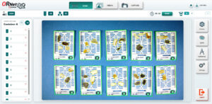
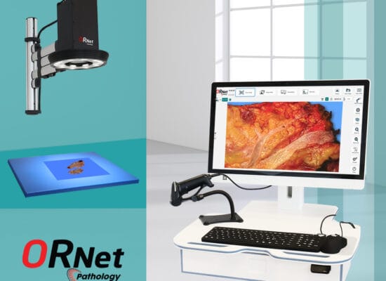
Document up to 12 cassettes in a single image — with automatic identification, tissue detection and measurement of every sample.
One image. Every cassette. Every fragment. Every measurement.
12 cassettes | 12 barcode reads | Unlimited sample analysis
Designed for biopsy material from different sources and tissue types, ORNet Biopsy Pathology creates a permanent digital record of the specimen while reducing manual documentation.
01 — From cassette to digital record
Automated documentation in seconds
Place up to 12 cassettes directly on the imaging surface. No special rack is required.
ORNet Biopsy Pathology automatically:
- IDENTIFIES
Reads and verifies the barcode/QR code of each cassette. - DETECTS
Recognizes every visible tissue fragment in the image — including very small samples. - MEASURES
Automatically determines the length, width and area of every detected tissue fragment. - CALCULATES
Provides the total number of fragments and total tissue area for each cassette. - VERIFIES
Checks the documented tissue area against laboratory-defined requirements and provides a warning when the documented quantity is insufficient. - DOCUMENTS
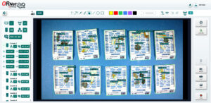
Creates a digital image containing the cassette information, tissue outlines, measurements and other relevant documentation.Up to 12 cassettes — documented simultaneously
One image can contain up to 12 cassettes. Each cassette is independently identified and documented. If additional cassettes are required, the same container can simply be used for the next batch.
Typical processing time: under 2 seconds, depending on network conditions.

Intelligent image analysis
Automated image recognition identifies tissue fragments individually, even when samples are very small.
If an area is not detected, the operator can use the correction tool to indicate the missed tissue directly on the image. ORNet Biopsy Pathology then reviews the selected area and incorporates it into the documentation and measurements.
02 — Every fragment. Every measurement. Permanently documented.See what was there before processing.
Small tissue samples can be difficult to document consistently using manual methods. ORNet Biopsy Pathology preserves a visual record of the specimen at the point of cassette documentation.Individual fragment analysis
Every detected tissue fragment is analyzed separately.
For each fragment, ORNet Biopsy Pathology can document:- Length / maximum cross-measurement
- Width
- Area
- Position within the cassette
- Visual image of the fragment
The system also calculates the total tissue area and total number of fragments for the cassette.

Laboratory-defined rules
Different specimen types may require different minimum quantities of tissue.
ORNet Biopsy Pathology allows laboratories to define their own rules and requirements.
The system automatically compares the documented tissue area or size with the configured requirement and alerts the user when the documented quantity is insufficient or sample too large.The image becomes part of the case record
The documented image can include:Cassette outlines + barcode/QR identification + individual tissue outlines + measurements + annotations
The result is a traceable visual record that can be stored, retrieved and integrated with other laboratory systems.Safer documentation
ORNet Biopsy Pathology supports safer specimen identification and documentation by connecting the physical cassette with its digital record.
From manual documentation to a traceable digital workflow.03 — Built for the digital pathology laboratory
Connect. Integrate. Scale.ORNet Biopsy Pathology is designed as a server-based solution, allowing deployment from a single workstation to large laboratories with multiple users and imaging stations.
LIS integration
Connect ORNet Biopsy Pathology to the laboratory information system using:API | HL7 | Other integration methods
Cassette identification and documentation can be connected directly to the appropriate case and specimen information.PACS & DICOM
Digital images can be stored locally, sent to LIS or integrated with existing imaging infrastructure using DICOM standard and sent to long-term storage.Flexible digital workflow
The digital documentation can be:- stored as part of the case record
- transferred to the LIS
- sent to PACS
- added to the documented image
- retrieved for later review
Scalable for every laboratory
Whether used at a single workstation or across a large pathology laboratory, the server-based architecture supports multiple users and multiple imaging stations.
ORNet Biopsy Pathology can grow with the laboratory.Towards the fully digital laboratory
ORNet Biopsy Pathology replaces manual and paper-based documentation steps with an automated, image-based digital workflow.
Less paper. Less manual work. More traceability

04 — Technology at a glance
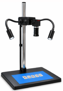
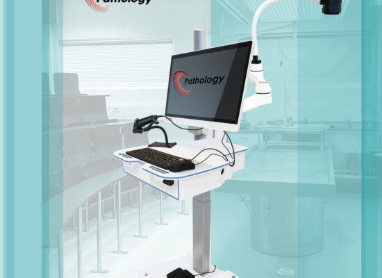
Intelligent imaging for small tissue samplesImaging with a mobile small-tissue documentation workstation:
4K / 48 MP USB camera
10× optical zoom
5–50 mm varifocal lens
Manual focus
Dedicated illumination — 2 × 150 lm lights
Designed to provide detailed, high-resolution images of tissue samples while maintaining natural-looking specimen colors as closely as possible.
Output formats
PNG / PDF / DICOM
Technical requirements:
User needs a Windows 11 PC with network capabilities.
Server with AI capable graphics card based on user quantity. Fast network.Documentation
Up to 12 cassettes per image- Automatic cassette detection
- Barcode / QR code reading
- Cassette verification
- Individual tissue-fragment detection
- Automatic measurement
- Length and width
- Individual fragment area
- Total tissue area
- Fragment count
- Image annotations
- Manual correction of detected areas
- Laboratory-defined verification rules
Designed for pathology
- Suitable for tissue samples from different biopsy sources
- Standard cassette workflow
- No dedicated cassette rack required
- Up to 12 cassettes can be documented simultaneously
- Server-based architecture
- Suitable for single-user and multi-user environments
- Scalable for large laboratories
Take the next step toward a fully digital, paper-free pathology laboratory.
Every sample documented. Every fragment accounted for. Every record digital.
ORNet Biopsy Pathology