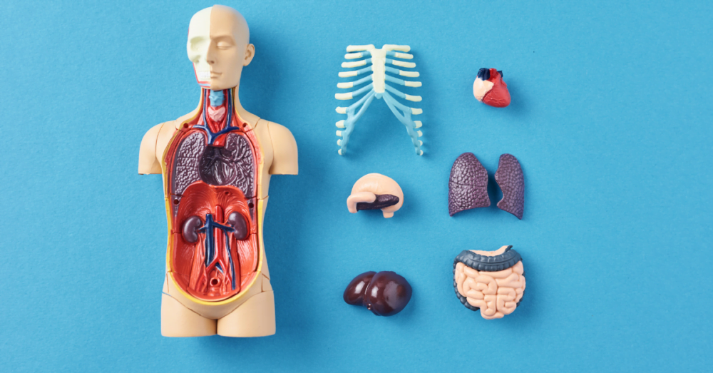
Gross Pathology, often considered the foundation of pathology, refers to the macroscopic assessment of pathology specimens. In other words, it is the study of diseases as they appear to the naked eye. There are two main aspects of it. First, examining organ tissues visually, without the use of a microscope, to identify abnormal changes that indicate disease. Secondly, preparation of the tissue for further microscopic examination.
Grossing acts as the first step in a diagnostic journey, providing the foundational analysis upon which further investigation builds. In a more general sense, grossing focuses on the identification of the most optimal part of the tissue sample for further examination.
Importance of Gross Pathology
From individual patient care to broader medical research, the importance of gross pathology remains an undeniable fact. It’s a testament to the adage, “Seeing is believing.” And in gross pathology, what we see forms the first crucial step in believing and understanding the story a disease tells.
When a patient presents with certain symptoms, clinicians have a multitude of potential diagnoses to consider. However, a gross examination of a biopsy or surgical specimen can provide invaluable clues that help narrow down these possibilities. Much like a sculptor first making broad strokes to outline a figure before adding detailed features, gross pathology provides the broad strokes that outline the nature of the disease.
Gross pathology can help determine the size, location, and extent of a disease; information crucial for formulating a treatment plan. It acts as a guide, assisting the clinician’s in their decision-making process. For instance, if a gross examination of a tumor specimen reveals it has invaded surrounding tissues, it can influence whether the patient needs further surgery or adjunctive treatments like chemotherapy.
Moreover, gross pathology’s importance extends beyond individual patient care. Findings from gross examinations contribute to our collective understanding of diseases. They form a part of medical databases and research studies, advancing the frontiers of medical science.
Types of Gross Pathological Examinations
There are different ways pathologists employ gross examination, depending on the specimen’s source: biopsies, surgical specimens, and autopsies.
Biopsies are small tissue samples taken from a living person to diagnose a disease or monitor its progression. In contrast, autopsies refer to the examination of a body after death, to determine the cause of death or study the disease’s extent and effects. Lastly, the surgical specimens are tissues removed during surgery. These tissues are examined to assess the disease and guide further treatment.
Gross Examination Procedure
The gross examination procedure is a methodical and meticulous process involving several crucial steps. It is an essential part of pathological examinations, providing a first-hand look at the specimen. Let’s break down each step for a better understanding.
Conducting a gross examination involves a systematic approach:
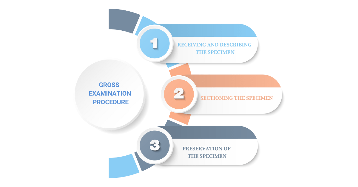
1. Receiving and Describing the Specimen
The initial step in the gross examination process is receiving and describing the specimen. The moment a specimen is received, its journey to reveal the secrets it holds begins.
The pathologist or a trained technician will document the specimen’s state upon arrival. Details such as the type of specimen, its source, the identity of the patient, and any clinical information provided are meticulously recorded.
Then the actual specimen is examined visually. Here, the pathologist notes its size, shape, color, texture, and any visible abnormalities. For instance, in the case of a tumor, the pathologist might record its size, its location, whether it’s solid or cystic, its color, and other surface characteristics.
This descriptive data, while may seem elementary, can be a treasure trove of initial clues about potential disease. Each detail could be a vital piece of the puzzle in the overall diagnosis process.
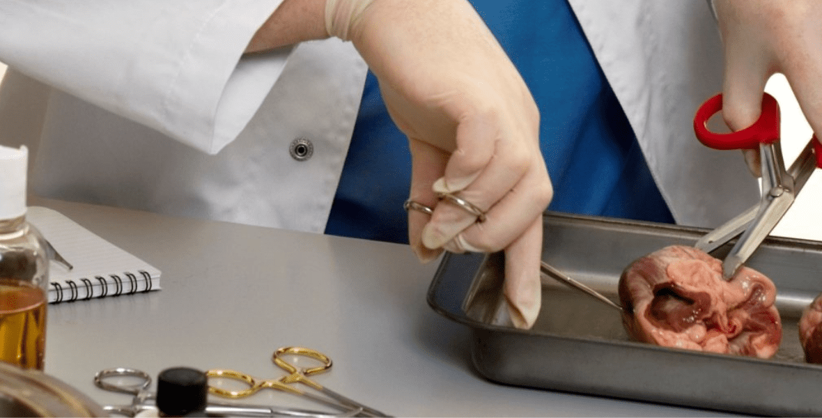
2. Sectioning the Specimen
Following the initial description, the specimen is then sectioned, or cut into thinner pieces. This is not random slicing but rather a precise process that depends on the type of tissue, its size, and the suspected disease.
Sectioning aims to reveal what’s inside the tissue. If we consider the example of a tumor again, sectioning would show the color of the cut surface, the consistency of the tissue (whether it’s hard, soft, or somewhere in between), and whether there are areas of hemorrhage or necrosis.
For some specimens, like certain types of tumors, a specific pattern of sectioning may be followed to ensure the pathologist can accurately assess the extent of the disease. Special techniques may be applied to trace the spread of the disease or to determine whether it has been completely removed.
3. Preservation of the Specimen
Once the gross examination is complete, the specimen doesn’t just get discarded. Instead, representative sections of it are preserved for potential further examination.
The specimen sections are typically placed in a solution of formalin, which serves to ‘fix’ the tissue, preserving its microscopic structure. These sections can then be processed further to create slides for microscopic examination if needed.
Moreover, in some cases, parts of the specimen may be stored in a different way for special tests. For instance, if there’s a need to look for certain proteins or genes in the tissue, the section may be snap-frozen and stored at very low temperatures.
The preservation of specimens plays a crucial role in maintaining a record of the patient’s disease. It provides an opportunity for further investigations and re-evaluations if needed later in the course of the patient’s disease management.
The Role of a Pathologist in Gross Examination
During the gross examination, a pathologist’s work involves a careful and critical analysis of the specimen, from its outer appearance to its internal structure. This meticulous approach ensures no significant detail is overlooked, which might be crucial in understanding the nature of the disease.
Their role begins with receiving and assessing the specimen. Their keen eye and experienced touch allow them to gauge the abnormal characteristics that might not be visible to an untrained observer. It is at this initial stage that they start forming a preliminary hypothesis about the disease.
The pathologist then proceeds to section the specimen, a process that often uncovers deeper insights about the disease. Like an expert geologist reading the layers of rock strata, the pathologist interprets the subtle variations in the tissue structure, discerning the story of the disease.
Their insights don’t stop at identifying the disease. The pathologist assesses the extent and severity of the disease, providing crucial data that directly impacts patient management. Their role extends beyond the laboratory, often collaborating with other clinicians to guide the optimal course of treatment.
Application of Gross Pathology in Medical Practice
The application of gross pathology in medical practice is vast and multifaceted. It is, in essence, the cornerstone on which many medical decisions are built.
At the heart of disease diagnosis, gross pathology provides critical insights. A tissue’s gross appearance can often provide immediate information about the presence and nature of a disease. This initial impression can guide further microscopic examinations and other diagnostic tests, ensuring a comprehensive and accurate diagnosis.
In the context of surgical interventions, gross pathology’s role is significant. By examining surgical specimens, pathologists can provide immediate feedback to surgeons regarding the disease’s extent and whether the surgical margins are clear of the disease. This real-time information can influence surgical decisions, potentially improving patient outcomes.
Gross pathology also plays a crucial role in post-mortem examinations or autopsies. It helps determine the cause of death and understand the disease’s progression and impact on the body. These findings can provide closure for the family, contribute to medical research, and even aid in public health planning.
Finally, gross pathology lays the groundwork for research into disease mechanisms. By understanding the gross changes in diseased tissues, scientists can develop hypotheses about the disease process, stimulating further research at the cellular and molecular levels.
In all these ways, gross pathology demonstrates its indispensable role in medical practice, enabling accurate diagnoses, guiding effective treatment, contributing to research, and ultimately, improving patient care.
Technological Innovations in Gross Pathology
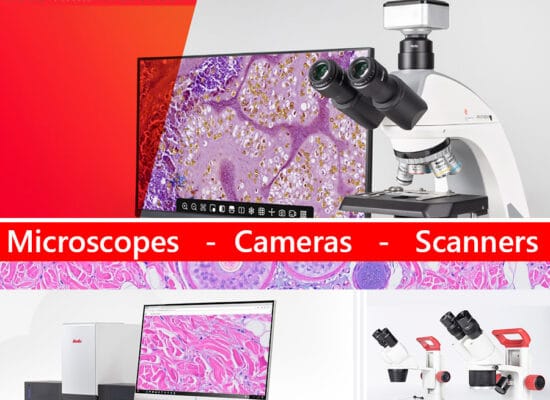
In the past, the tools of a pathologist were relatively simple – a keen eye, a steady hand, and a sharp scalpel. However, the landscape of gross pathology is no longer what it used to be, having been significantly influenced and enhanced by technological innovations. These advancements have revolutionized the field, enriching our understanding of diseases and refining our diagnostic capabilities.
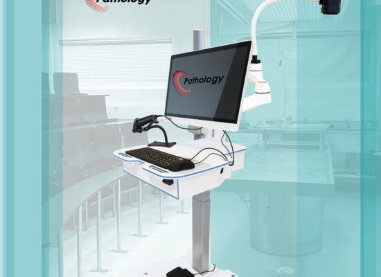
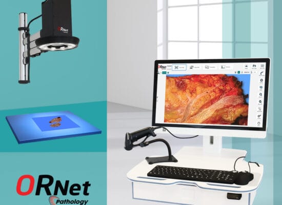
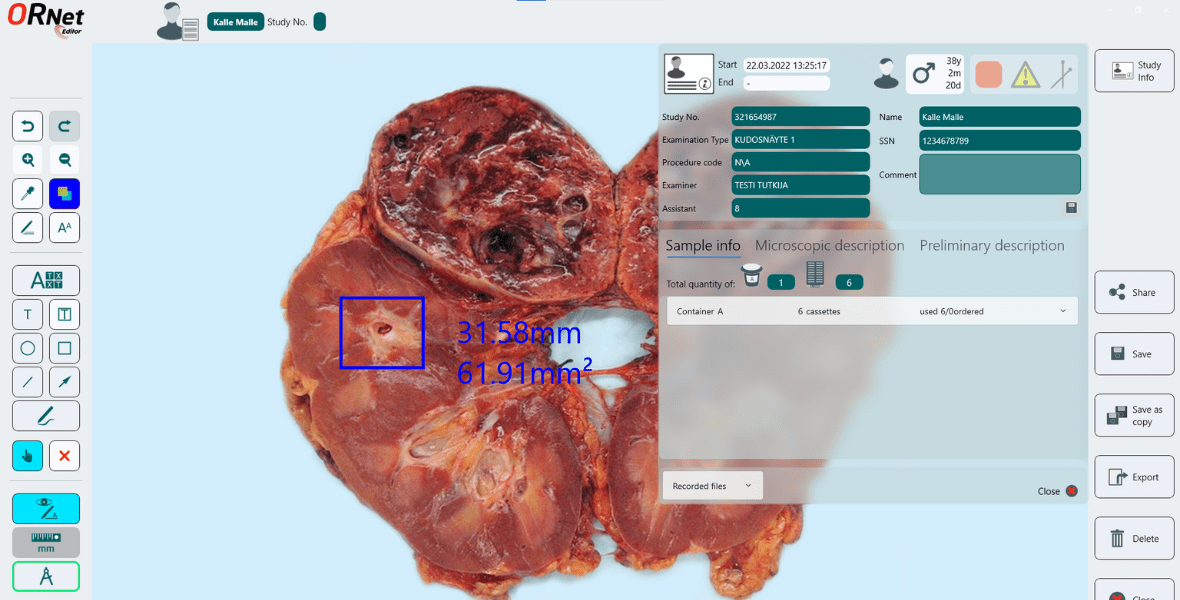
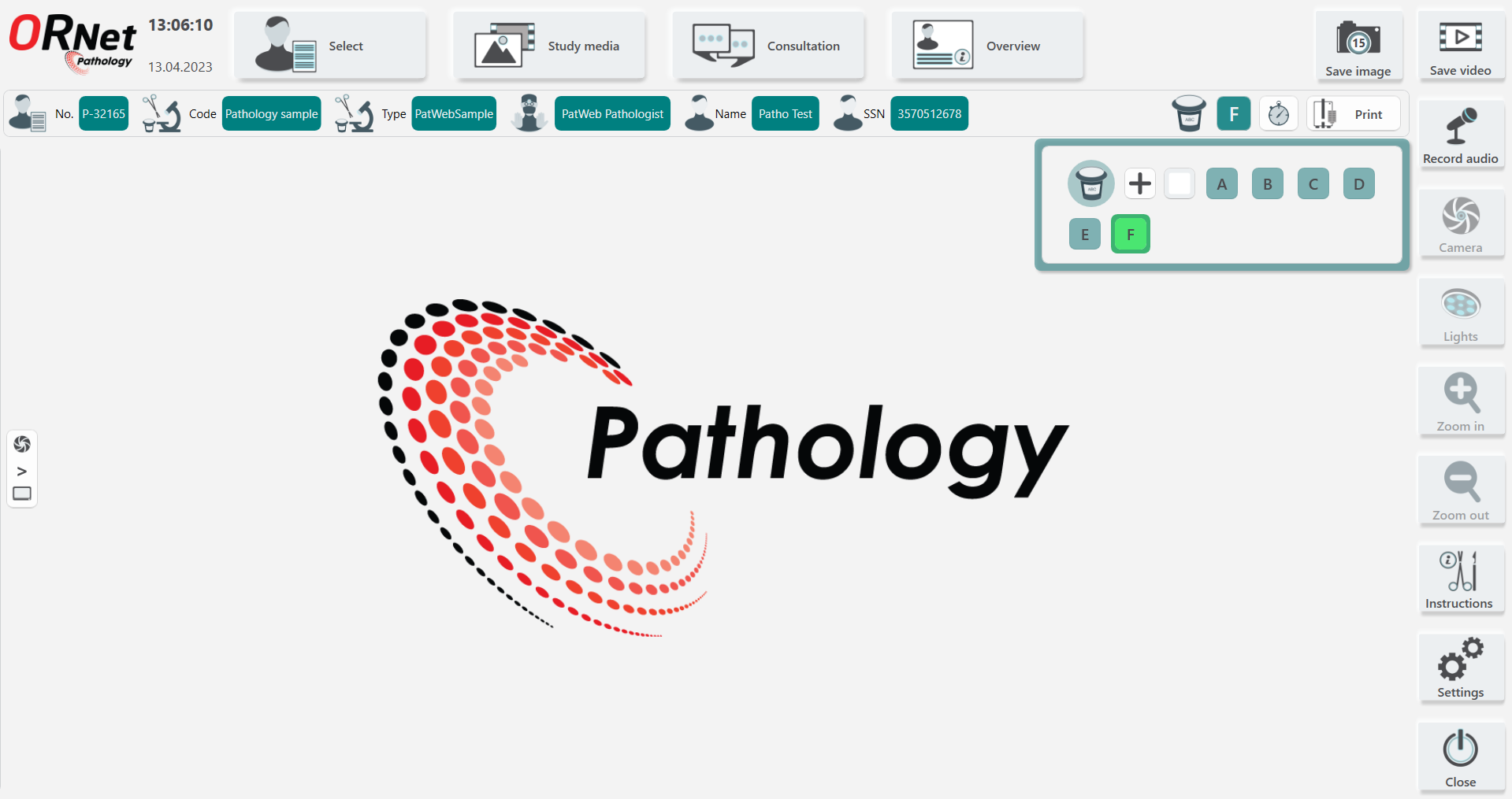
One of the most impactful innovations is the advent of Digital Pathology. This involves capturing high-resolution images of specimens, enabling pathologists to examine and analyze tissues digitally. This technology brings several advantages, such as the ability to zoom in on specific areas, share images for collaborative analysis, and store images for future reference. In this regard, tools like ORNet Pathology are paving the way with comprehensive capabilities and ease of adoption.
Another noteworthy innovation is 3D Imaging and Reconstruction. This technology allows pathologists to visualize specimens in three dimensions, providing a more comprehensive understanding of the specimen’s structure. It’s especially beneficial in complex cases where 2D images might not capture the full extent of the disease.
Moreover, Artificial Intelligence (AI) is making significant strides in gross pathology. Machine learning algorithms can analyze digital images of specimens, identify patterns that might be overlooked by the human eye, and even predict certain disease characteristics. While AI does not replace human expertise, it serves as a powerful tool to aid and enhance the pathologist’s diagnostic abilities.
Telepathology, the practice of pathology at a long distance, is another breakthrough in the field. It allows digital images to be transmitted remotely, enabling pathologists to consult with colleagues around the world. This fosters collaborative analysis, which can be particularly beneficial in challenging cases.
Moreover, advancements have been made towards Automated Specimen Processing, with some devices now capable of measuring specimen dimensions or capturing gross images automatically. While full automation of grossing is not yet available, these steps towards automation are promising, aiming to increase precision and decrease manual errors in specimen processing.
These technological advancements, and many more, are propelling gross pathology into the future. By enhancing the detail and accuracy of gross examination, they are reinforcing gross pathology’s crucial role in disease diagnosis and treatment, and paving the way for exciting new developments in the field.
Conclusion
In essence, gross pathology serves as the first significant step in unveiling the mysteries hidden within tissues and cells, setting the foundation for disease diagnosis and treatment planning. Its importance cannot be overstated, and with ongoing technological advancements, the future of gross pathology holds even more promise.
FAQs
Grossing in pathology means describing and measuring the color, size, weight, etc of the tissue sample, inking it to highlight different areas, and sectioning it for further analysis.
A gross pathologist studies disease as it appears to the naked eye, usually through the examination of organs and tissues. They perform macroscopic analysis on the specimen and prepare tissues for microscopic examination.
Gross pathology is the first step and plays a critical role in guiding accurate diagnoses and subsequent treatment plans. It provides immediate clues about a disease’s nature and severity.
Some examples of gross examinations include autopsies, surgical specimens, and biopsies.